A chronic disorder of carbohydrate metabolism due to relative or absolute insulin deficiency. Most cases of spontaneous diabetes occur in middle‐aged dogs and middle‐aged to older cats. In dogs, females are affected about twice as often as males, and certain small breeds (for example, Miniature Poodles, Dachshunds, Schnauzers, Cairn Terriers, and Beagles) show increased incidence—though any breed may be affected. In cats, one study found obese males were more commonly affected than females; no clear breed predilection is identified in cats.
Etiology and Pathogenesis
The pathogenic mechanisms responsible for reduced insulin production and secretion are multiple. Typically, they relate to islet-cell destruction or dysfunction: in dogs this is often secondary to immune destruction or severe pancreatitis; in cats to amyloidosis of the islets. In dogs, chronic relapsing pancreatitis causes progressive loss of both exocrine and endocrine pancreatic cells and replacement by fibrous connective tissue, eventually producing a firm, multinodular pancreas which may contain areas of necrosis and haemorrhage. Over time the pancreas may contract so that only a thin fibrous band remains near the duodenum and stomach. In other cases the number of β-cells decreases and the remaining cells become vacuolated; in long‐standing disease the islets may become hard to identify. Insulin resistance and secondary diabetes mellitus are also observed in many dogs with spontaneous hyperadrenocorticism or after long-term administration of glucocorticoids or progestins. Pregnancy and the diestrus phase may also predispose to diabetes mellitus. In dogs (but not cats), progesterone induces growth hormone release from mammary tissue, leading to hyperglycaemia and insulin resistance. Obesity also predisposes to insulin resistance in both dogs and cats.
In cats with diabetes mellitus, distinctive degenerative lesions are usually found, localized selectively in the islets of Langerhans while the remainder of the pancreas appears normal. The most common lesion is the selective deposition of amyloid (derived from islet‐associated polypeptide, IAPP, which is co-secreted with insulin) in the islets, resulting in β‐cell degeneration. Cats cannot process IAPP properly, leading to its accumulation, conversion into amyloid, and eventual disruption of β‐cell function and insulin resistance. As cats age, a larger proportion of their islets contain amyloid; diabetic cats have more islets affected and greater amyloid accumulation compared with age-matched non-diabetic cats (amyloid + IAPP → β-cell disruption → insulin resistance → diabetes).
Infections with certain viruses can cause selective islet damage or pancreatitis in humans, and have been speculated as causes of rapidly‐onset diabetes mellitus, though this has not yet been documented in dogs or cats. In such cases of β-cell degeneration/necrosis there is often infiltration of islets by lymphocytes and macrophages. Stress, obesity and corticosteroid or progestogen administration may increase severity of clinical signs.
The full expression of the complex metabolic derangements in diabetes mellitus appears to involve a bihormonal abnormality. While insulin deficiency (absolute or relative) in response to rising extracellular glucose has long been recognised as the major hormonal abnormality, recent emphasis has been placed on the importance of an absolute or relative increase in glucagon secretion. Hyperglucagonemia may result from increased pancreatic glucagon release, increased enteroglucagon, or both. Elevated glucagon contributes to severe hyperglycaemia by mobilising hepatic glucose stores and contributes to ketoacidosis by increasing hepatic fatty acid oxidation.
Clinical Findings
Onset of diabetes is often insidious and the clinical course chronic. In dogs, common signs include polydipsia, polyuria, polyphagia combined with weight loss, bilateral cataracts, and weakness. The disturbances in water metabolism develop primarily because of osmotic diuresis. The renal threshold for glucose is about ~180 mg/dL in dogs and ~280 mg/dL in cats. Merck Veterinary Manual+1
Diabetic animals have reduced resistance to bacterial and fungal infections and often develop chronic or recurrent infections such as cystitis, prostatitis, bronchopneumonia and dermatitis. This susceptibility may be partially due to impaired neutrophil function (chemotaxis, phagocytosis, antimicrobial activity). Radiographic evidence of emphysematous cystitis (although rare) — caused by glucose‐fermenting organisms such as Proteus sp., Aerobacter aerogenes, and Escherichia coli resulting in gas formation in the bladder wall and lumen — is suggestive of diabetes mellitus. Emphysema may also develop in the gallbladder wall in diabetic dogs. Merck Veterinary Manual
Hepatomegaly due to lipid accumulation is common in diabetic dogs and cats. Fatty liver occurs as a consequence of increased fat mobilization from adipose tissue. Individual hepatocytes become greatly enlarged by multiple neutral‐lipid droplets. In cats, hepatic lipidosis may occur in conjunction with diabetes mellitus. Merck Veterinary Manual+1
Cataract formation occurs frequently in dogs (but rarely in cats) with poorly‐controlled diabetes mellitus. In dogs the lenticular opacities initially appear along suture-lines of lens fibres and are stellate (“asteroid”) in shape. Cataract formation in dogs is related to the unique sorbitol pathway of glucose metabolism in the lens (leading to lens edema and disruption of light transmission). Although the sorbitol pathway appears present in cats, cataract development is rare in them. Other extrapancreatic lesions seen in humans (e.g., nephropathy, retinopathy, micro- and macrovascular angiopathy) are rare in dogs and cats. Merck Veterinary Manual
Diagnosis
Diagnosis of diabetes mellitus is based on persistent fasting hyperglycaemia and glycosuria. The normal fasting blood-glucose in dogs and cats is 75–120 mg/dL. In cats, stress-induced hyperglycaemia is a common problem and multiple blood and urine samples may be required to confirm diagnosis. The measurement of serum fructosamine can help differentiate stress-induced hyperglycaemia (where fructosamine is normal) from true diabetes mellitus. In all cases, a search should be made for drugs or diseases predisposing to diabetes. PubMed+2Merck Veterinary Manual+2
Treatment
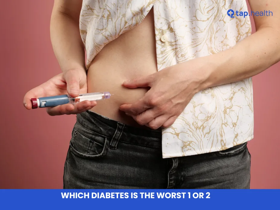
Long‐term success in management depends on owner understanding and cooperation. Treatment involves a combination of weight reduction, dietary management, insulin therapy, and possibly oral hypoglycaemics. Intact females should be spayed/ neutered. In cats, recent evidence supports use of high-protein, low-carbohydrate diets. In dogs, diets high in fibre and complex carbohydrates are preferred. Dietary and weight‐reduction alone will not control the disease; thus initial therapy with insulin is required. In most dogs two doses of insulin daily are necessary. Generally, NPH or lente insulin is used initially at ~0.5 U/kg, twice daily. With twice‐daily injections, two meals of equal calorie content are given at the time of insulin administration. Avoid diets high in simple sugars (semi-moist foods). Clinical signs plus serial blood‐glucose determinations are used to monitor therapy after initial stabilisation at home for 5–7 days.
In dogs failing to achieve good glycaemic control on NPH or lente insulin, basal insulin detemir may be considered; because of its potency, the starting dose is ~0.1 U/kg twice daily, with reassessment of clinical signs and glycaemic control after 1 week.
Home blood‐glucose monitoring is preferable to avoid changes in the pet’s routine and stress of in-hospital testing. Studies in both dogs and cats demonstrate that at-home monitoring improves glycaemic control and increases the likelihood of remission in diabetic cats. In cats, a regimen of insulin glargine plus high-protein, low-carbohydrate diet is associated with remission of diabetes and discontinuation of insulin therapy in ~80-90% of cases within the first 3–4 months of treatment. NPH, lente or PZI insulins may also be used in cats, with starting doses of 1-3 units twice daily; however these insulins are not associated with high remission rates. The oral hypoglycaemic agent glipizide (a sulfonylurea) has been evaluated in diabetic cats: initial dose ~2.5 mg twice daily PO, plus dietary management. Clinical response may be seen in 3–4 weeks. Short-term success is seen in ~50% of treated cats; long‐term success (>1 yr) is ~15%. Alternatives glimepiride (2 mg/day) or glyburide (0.625 mg/day) may also be used in cats. Acarbose (an α-glucosidase inhibitor) has been used in cats at ~12.5-25 mg twice- or three-times daily, in conjunction with diet and/or insulin to control hyperglycaemia.
Ketoacidosis is a serious complication and is a medical emergency. Therapy includes correcting dehydration by IV fluids (0.9% NaCl or Lactated Ringer’s), reducing hyperglycaemia and ketosis by administration of crystalline zinc (regular) insulin, maintaining serum electrolyte levels (especially potassium) via supplemental electrolyte solutions, and identifying and treating underlying/ complicating diseases such as acute pancreatitis or infections. Numerous insulin regimens are used in ketoacidotic diabetes mellitus. In the intermittent insulin regimen: regular insulin at ~0.2 U/kg IM initially, followed by hourly 0.1 U/kg; once serum glucose is <250 mg/dL, insulin is administered SC at ~0.25-0.5 U/kg every 4-6 hr, with careful serum-glucose monitoring at 1-2 hr intervals. During aggressive insulin therapy, blood glucose may fall rapidly, and addition of 2.5–5% dextrose to IV fluids may be required.
Once insulin therapy has been instituted, glucose should be checked frequently until an adequate maintenance dose is determined. Once the patient is on maintenance therapy and stable, reassessment is recommended every 4-6 months. VetSmall
External Links
- “What’s in a Name? Classification of Diabetes Mellitus in Veterinary Medicine” – PMC article. Link PMC+1
- “Nutritional Management of Cats and Dogs with Diabetes Mellitus” – PubMed article. Link PubMed
- “2018 AAHA Diabetes Management Guidelines for Dogs and Cats” (PDF) – Association guidelines. Link AAHA
- “Insulins for the Long Term Management of Diabetes Mellitus in Dogs” – BMC review. Link BioMed Central
- “Diabetes Mellitus in Dogs and Cats” – Merck Veterinary Manual online. Link