Table of Contents
- Imaging Techniques for Liver, Pancreas & Fat in Diabetes
- How Imaging Helps Diagnose Diabetes-Related Organ Damage
- Liver, Pancreas, and Fat Assessment: Key Imaging Findings in Diabetes
- A Guide to Imaging in Diabetes: Understanding Liver, Pancreas, and Fat
- What Imaging Tests Reveal About Diabetes’ Impact on Organs?
- Frequently Asked Questions
- References
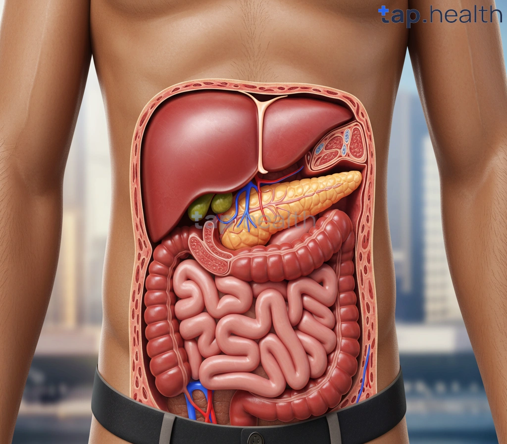
Diabetes management often relies on understanding more than just blood sugar levels. Understanding Liver, Pancreas, and Fat: Imaging in Diabetes plays a crucial role in diagnosing and monitoring this complex disease. This blog post will explore how advanced imaging techniques provide invaluable insights into the health of these vital organs, which are central to glucose metabolism. We’ll delve into the different imaging modalities used, their benefits, and limitations, helping you better understand your own health or the health of your patients. Let’s unravel the mysteries behind these internal organs and their impact on diabetes management.
Imaging Techniques for Liver, Pancreas & Fat in Diabetes
Assessing Organ Health in Diabetes
Diabetes significantly impacts liver, pancreas, and fat distribution, increasing the risk of complications like fatty liver disease (NAFLD), pancreatitis, and cardiovascular issues. Given that 61% of people with diabetes are aged between 20-64 years, early detection is crucial in India and tropical countries where diabetes prevalence is high. Imaging plays a vital role in assessing the health of these organs and guiding treatment strategies.
Key Imaging Modalities
Several imaging techniques provide valuable information. Ultrasound is a readily available, cost-effective first-line approach for evaluating liver size, texture, and the presence of fatty infiltration. Computed tomography (CT) scans offer detailed anatomical images, useful for detecting pancreatic abnormalities and assessing fat distribution. Magnetic resonance imaging (MRI), including advanced techniques like MR elastography, provides more precise assessment of liver fibrosis and fat content. In specific cases, endoscopic ultrasound (EUS) may be employed for detailed visualization of the pancreas. The choice of imaging modality depends on the clinical presentation, accessibility of technology, and individual patient needs, particularly important in resource-constrained settings common in many tropical regions.
Regional Considerations
Access to advanced imaging modalities like MRI may be limited in some parts of India and other tropical countries. Therefore, strategies focusing on cost-effective initial screening methods like ultrasound, coupled with targeted use of CT or MRI based on clinical suspicion, are vital. Moreover, awareness campaigns emphasizing early detection and management of diabetes, particularly within the 39% of the diabetic population aged 65+, are crucial for improving outcomes. Understanding the link between diabetes and organ health is paramount, and learning more about The Link Between Diabetes and Fatty Liver can be beneficial.
Actionable Steps
Regular check-ups with your physician are essential for individuals with diabetes. Open communication regarding any symptoms or concerns related to liver, pancreas, or unexplained weight changes is crucial for early intervention and improved management of diabetes and its complications. Discuss appropriate imaging options with your doctor to determine the best approach for your specific situation. Furthermore, exploring How Can New Technological Advances Improve Diabetes Lifestyle? can empower individuals to better manage their condition.
How Imaging Helps Diagnose Diabetes-Related Organ Damage
Diabetes significantly impacts various organs, often silently progressing until complications arise. Early detection is crucial, especially in high-risk populations prevalent in Indian and tropical countries. Imaging techniques play a vital role in identifying and monitoring this organ damage, improving patient outcomes and reducing long-term complications.
Liver and Pancreas Imaging in Diabetes
Fatty liver disease (FLD), frequently associated with type 2 diabetes, can be detected using ultrasound, CT scans, or MRI. These imaging modalities assess liver size, texture, and the presence of fat accumulation. Similarly, imaging helps evaluate the pancreas for signs of inflammation or fibrosis, common in diabetic patients. Early identification through these techniques allows for timely interventions, potentially preventing further progression to more serious conditions.
Assessing Diabetic Nephropathy and Cardiovascular Issues
Advanced diabetes can lead to kidney damage (diabetic nephropathy). Ultrasound or CT scans can help assess kidney size and function. Furthermore, imaging plays a critical role in assessing cardiovascular complications, a leading cause of mortality in diabetics. CT angiography and echocardiography can identify coronary artery disease, heart failure, and other cardiovascular issues. Understanding How Does Diabetes Affect Blood Flow? is crucial to comprehending the cardiovascular risks.
Importance of Foot Imaging and Prevention of Amputations
The risk of foot ulcers is a significant concern for diabetics, with nearly 15% experiencing them in their lifetime, leading to high amputation risks. Early detection of foot problems is crucial. Imaging techniques like X-rays and MRI can identify underlying bone or soft tissue damage, guiding early intervention and potentially preventing the need for amputation. Regular foot checks and prompt medical attention are paramount, particularly for individuals in regions with limited access to advanced healthcare.
Conclusion: Accessing Timely Care
Early diagnosis through imaging is vital for managing diabetes and its complications, particularly in Indian and tropical countries. Seeking regular check-ups, including imaging assessments as recommended by your physician, is crucial for preventing devastating complications like limb amputations and improving your overall quality of life. Don’t delay; prioritize your health and seek timely medical attention. For more on leveraging technology for better diabetes management, read about How AI Helps in Monitoring and Managing Diabetes.
Liver, Pancreas, and Fat Assessment: Key Imaging Findings in Diabetes
The Crucial Role of Imaging in Diabetes Management
Diabetes significantly impacts multiple organs, and early detection is vital for effective management. The alarming statistic that nearly 30% of diabetics develop diabetic nephropathy highlights the importance of proactive healthcare. Imaging plays a crucial role in assessing the health of the liver, pancreas, and fat tissues—organs frequently affected in diabetes. Changes in these organs can indicate complications like Fatty Liver disease (often prevalent in Indian and tropical countries), pancreatitis, and insulin resistance. Regular imaging helps in early diagnosis and timely intervention.
Imaging Modalities and Key Findings
Ultrasound, CT scans, and MRI are commonly used imaging techniques to assess these organs. Ultrasound is often the first-line investigation due to its accessibility and cost-effectiveness, particularly beneficial in resource-constrained settings prevalent across many Indian and tropical regions. In the liver, imaging helps detect fatty infiltration, cirrhosis, and other liver-related complications. For the pancreas, imaging identifies inflammation, cysts, or tumors associated with diabetes. Finally, imaging aids in assessing the distribution and quantity of visceral fat, a significant contributor to insulin resistance and metabolic syndrome. Abnormal findings in these organs often warrant further investigation and tailored management strategies.
Actionable Steps for Better Diabetes Care
Regular check-ups with a physician are crucial. Open communication with your doctor about your family history, lifestyle, and any symptoms is essential. Advocating for appropriate imaging tests based on your risk profile and discussing the findings with your healthcare team will ensure the best possible management of your diabetes. Early detection and intervention facilitated by regular imaging significantly improve patient outcomes and quality of life. Remember, proactive healthcare is key to managing diabetes effectively, especially in the context of prevalent comorbidities in Indian and tropical populations. Understanding the connection between diabetes and weight management is also crucial, as highlighted in Understanding the Link Between Diabetes and Obesity.
A Guide to Imaging in Diabetes: Understanding Liver, Pancreas, and Fat
The alarming statistic that 50% of diabetes cases worldwide remain undiagnosed, as highlighted by the International Diabetes Federation, underscores the critical need for early detection and management, particularly in high-prevalence regions like India and other tropical countries. Imaging plays a crucial role in this process, offering valuable insights into the health of vital organs often affected by diabetes: the liver, pancreas, and fat deposits.
Liver Imaging in Diabetes
Diabetes can lead to non-alcoholic fatty liver disease (NAFLD), a condition characterized by excessive fat accumulation in the liver. Imaging techniques like ultrasound, CT scans, and MRI can detect and assess the severity of NAFLD, allowing for timely intervention and management of associated complications. Early detection is especially important in Indian and tropical populations, where NAFLD prevalence is high.
Pancreas Imaging in Diabetes
The pancreas, responsible for insulin production, is central to diabetes. Imaging techniques can help identify pancreatic abnormalities that might contribute to diabetes development or its complications. MRI and CT scans are particularly useful in evaluating pancreatic structure and function, aiding in diagnosis and treatment planning.
Fat Assessment in Diabetes
Abdominal fat accumulation is strongly linked to insulin resistance and diabetes. Imaging modalities can quantify visceral fat (deep abdominal fat), helping clinicians assess metabolic risk and tailor treatment strategies. In tropical climates, understanding the impact of lifestyle factors on abdominal fat distribution is crucial for effective diabetes management. Managing diabetes effectively becomes even more critical as we age, and understanding the specific challenges at each life stage is important. For more information, see our guide on Managing Diabetes as You Age: Challenges and Solutions.
Conclusion
Regular health check-ups, including appropriate imaging where indicated, are crucial for individuals in India and other tropical countries, particularly those with a family history of diabetes or associated risk factors. Early detection through imaging can significantly improve diabetes management, preventing or delaying serious complications and improving quality of life. Consult with your doctor to discuss the appropriate imaging techniques for your individual needs. Protecting your vision is also a key aspect of long-term diabetes management. Learn more about essential eye care tips in our article, How to Protect Your Vision with Diabetes: Essential Eye Care Tips.
What Imaging Tests Reveal About Diabetes’ Impact on Organs?
Diabetes significantly impacts major organs, contributing to the staggering $327 billion annual cost in the US alone. This burden is felt acutely in India and other tropical countries, where diabetes prevalence is rising. Understanding how diabetes affects the liver, pancreas, and fat is crucial for timely intervention and better management.
Imaging the Liver: Detecting Fatty Liver Disease
Non-alcoholic fatty liver disease (NAFLD) is a common complication of diabetes. Ultrasound, CT scans, and MRI can detect fat accumulation in the liver, assessing the severity of NAFLD and its potential progression to cirrhosis. Early detection through these imaging techniques is vital in high-risk populations, especially in regions with high rates of diabetes like India. Regular checkups are strongly recommended for individuals with diabetes residing in tropical countries.
Pancreas Imaging: Assessing Function and Damage
The pancreas plays a central role in blood sugar regulation. Imaging techniques like CT scans and MRI can assess pancreatic structure and function, helping to identify issues such as pancreatitis or pancreatic cancer, which have a higher incidence in individuals with long-standing diabetes. These imaging modalities help in early diagnosis and treatment planning, improving patient outcomes.
Imaging Abdominal Fat: Understanding Metabolic Risk
Abdominal fat accumulation, particularly visceral fat, is strongly linked to diabetes complications. CT scans and MRI can precisely quantify visceral fat, helping assess metabolic risk and guide lifestyle interventions. This is particularly relevant in tropical regions where dietary patterns and lifestyle factors can contribute to increased abdominal fat. Early intervention, focusing on diet and exercise, is crucial in mitigating these risks. It’s also important to be aware of how diabetes can affect other systems, such as the respiratory system. For more information, read our article on How Does Diabetes Affect the Respiratory System?
Conclusion: Proactive Healthcare in Tropical Regions
Regular health checkups, including appropriate imaging tests, are essential for individuals with diabetes in India and other tropical countries. Early detection and management of organ damage can significantly improve quality of life and reduce the long-term health burden. Consult your doctor to discuss the suitability of imaging tests based on your individual risk factors and medical history. The link between diabetes and cancer is also a significant concern. To learn more, please see our blog post on Does Diabetes Cause Cancer?
Frequently Asked Questions on Understanding Liver, Pancreas, and Fat
Q1. How does diabetes affect my liver, pancreas, and fat distribution?
Diabetes can lead to fatty liver disease, pancreatitis, and unhealthy fat distribution, increasing your risk of serious health problems.
Q2. Why is early detection of diabetes-related complications so important?
Early detection allows for timely intervention, preventing or delaying serious complications like cardiovascular issues and improving your quality of life.
Q3. What imaging techniques can help assess the health of my liver, pancreas, and fat?
Ultrasound is a cost-effective initial screening method. CT and MRI scans, including MR elastography, offer more detailed information.
Q4. How can I manage diabetes and its potential complications effectively?
Regular check-ups with your physician, open communication about your health, and prompt treatment of any issues are crucial for managing diabetes and improving outcomes.
Q5. What if advanced imaging techniques like MRI aren’t readily available in my area?
A tiered approach is often used. Ultrasound may be the initial screening tool, with more advanced imaging reserved for cases with higher clinical suspicion.
References
- A Practical Guide to Integrated Type 2 Diabetes Care: https://www.hse.ie/eng/services/list/2/primarycare/east-coast-diabetes-service/management-of-type-2-diabetes/diabetes-and-pregnancy/icgp-guide-to-integrated-type-2.pdf
- Diabetes Mellitus: Understanding the Disease, Its Diagnosis, and Management Strategies in Present Scenario: https://www.ajol.info/index.php/ajbr/article/view/283152/266731
Meta Data
Meta Title: Liver, Pancreas & Fat Imaging in Diabetes | Essential Guide
Meta Description: Discover the critical role of imaging in diagnosing diabetes. Learn how scans of your liver, pancreas, and fat tissue help manage and prevent diabetes complications. Read now!
alt_text: Liver, pancreas, and fat tissue medical scan images
url_slug: diabetes-organ-imaging